About ossteoarthritis and its causes,symptoms and treatment
By osteoarthritis, there is gradual degeneration and destruction of the cartilage at the bone ends and changes in the synovial mambrane, causing movement problems and pain.
The destruction of the cartilages can get so total that the bone ends get in contact with each other and wear each other further down.
The condition affects many persons as they grow older, but younger persons that use their joints heavily may also be affected. The condition mostly affect joints that have to sustain heavily load, for example the knee joints and hip joints, but any joint can be affected, including the spine. The hands and feet are often affected.
By the age of 65 80% of the population have signs of osteoarthritis by radigraphic exploration, but many will not have external symptoms.
Symptoms of osteoarthritis
The symptoms of osteoarthritis is pain by movement. Graually the joints also do not bend as smoothely as before, as if the joint are filled with sand. There can be cracking noises by movement of the joint. Muscular weakness can occur.
There is often excessive fluid in the joint, but this fluid is not normal synovial fluid. The affected joint is often swallen, but not to the extend that one can see by rheumatoid arthritis. The joint tend to feel worse the more it is used during the day, and this is contrary to what happens with rheumatoid arthritis.
In severe cases the joint can be totally locked.
The causes and mechanisms of osteoarthritis
Heavy use and prolonged mechanical stress at the joints over long time is a causal factor for osteoarthritis, the same is trauma to the joint.
Gradually the cartlage is worn away, and bare bone ends will appear and meet each other, first in the circumference of the bone ends, but later also the cartilage in the middle can be erased. In severe cases the bone ends also wear further down, and one bone can be locked inside the other.
There is usually no marked inflammation by osteoarthritis in the initial stages, but the damages can trigger inflammations.
There will be regrowth of the cartilage and the destructed bones to some extend, but the regrowth can produce abnormal structures and make the problem worse. Inflammation caused by reaction of the immune system to the damages can also make the destruction and problem worse.
The bare bone ends can react by producing a hard ivory-like bone mass as a means to remend the problem. Because the affected joint is used less and get a decreased range of movement, muscles connected to the joint can degenerate.
The primary causes of osteoarthritis
But all people using their body and joints in a demanding way do not get worn joints or orteoarthritis, and if they get it, not to the same degeree. Therefore there must in addition be some kind of weakness that makes the joints more woulnerable to mechanical wearing.
The mechanical strength of the cartilage may be decreased by many cases of osteoarthritis.
There may be some decreased repair of the wearing that occures daily so that the linings of the bone ends are gradually worn down or.
There may be some problems with the synovial fluid that lubricates the cartilage ends. A low level of glucosaminoglycans in the synovial fluid seems to give more friction inside the joint, and contribute to the erasing process. A decreased secretion of synovial fluid also may contribute to the process.
Allergic reactions may be a component in the cource.
Infection with fungi or other microbes ijn the joints may also be a causal factor. The infection makes the tissue in th ebone end weaker.
A history of frequent joint injury can weaken the cartilage tissue so that osteoarthritis more easily develops. This often happen to athletes like bascetball pålayers, football players, soccer players or dancers.
Lack of exercize is as heavy a causal factor for osteoarthritis like over-use. Exercize stimulate the healing processes in the joints.
Over-weight or obesity contribute as a causal factor for osteoarthritis, especially in the weight-carrying joints like th ehip and knees.
Probably malnutrition contribute to the abnomalities that make the joints more woulnerabel to daily wearing.
The tendency to get osteoarthritis seem to be inherited. That means that some mechanical weakness in the catilages or in the bones can be inherited. Also a decreased ability to repair tissue damage after injury and wearing may be inherited. Furthermore there may be an inherited weakness in the production of synovial fluid.
Osteoarthritis is to some extend regarded as part of the normal aging process.
Treatment for osteoarthritis
There is no good traditional medical treatment for ostoartritis. In severe cases urgery to change injured components of the joints is performed. Pain medication and non-steroid anti-inflammatory agents are used as treatments.
A ballanced mixture of active movement or exercises and rest of the affected body parts is beneficial. Avoiding exercises alltogether will usually worsen the condition, but the exercises should not make a very strong impact on the affected joints.
Swimming is a good exercise that give little impact on the joints but good muscular and condition training. Local heat before, and cold packs after exercise, can help relieve pain and inflammation.
For over-weight persons, a successful weight-loss program will greatly help against the condition or help tha afflicted persons to compensate for the problems.
Movement and posture aids like knee braces, walking cane, back brace or a walker can reduce pressure and ease walking.
Althernative medicines have the aim of stimulating the cartilage and bone to regrow, and to furnish nutrients for this regrowth. They also have the aim of improving the quality of the synovial fluid lubricating the insides of the joints, and thus decrease friction and prevent injury of the inside structures of the joints.
About joints
Joints are connections between two or more bones that can be bent in specific
directions depending on the anatomy of the joint. There are three types of
joint: Fibrous joint, cartilagious joint and synovaial joint.
Morphology of fibrous joints
In a fibrous joint two bones are connected just by fibrous connective tissue. These
types of joint have little mobility. They are found for example between the
bones in the scull. The small mobility of the joints gives the some elasticy an
high rigidity. The rigidity protects against compression and the
elasticity secures that the bones are not so easily crushed.
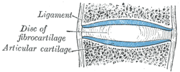
Morphology of cartilaginous
joints
In these type of joints each bone end have hard cartilage plate. Between the plates
of hard carttilage there is a disk of fibrous elastic fissue. In the middle
of the disk there is some fluid stuff. Around the joint there are strong fibrous ligaments with some elasticity. These kind of joints are found between the
vertebras in the spine. Such joints are rigid, but also very elastic so that
they can be bent fairly much in every direction when enough force is used.
Anatomy of a cartilagious joint (A free picture from wikipedia.org)
Morphology of synovial joints
In these joints the bone ends are seprated by a narrow space filled with
synovial fluid that lubricate the joint.
The bone ends are covered by a plate of hard cartilage. One end is usually
vonvex and the other concave and are fit to each other in size and shape. The
joint is surrounded by a fibrous joint capsule and at the outside there are
strong ligaments too. The inner lining of the capsule is a mucosa that produces
the synovial fluid, called synnovial membrane.
In many joints the capsule and synovial membrane forms slime bags called bursae.
A bursa increases the production of synovial fluid and also work as a pad to
buffer against impacts. In some joints
are part of the cave is divided into two by a cartilage disc, joint disc.
The knee joint also have a component called meniscus. There are one meniscus at
each side of the knee joint. The menischi are conical pads of carilage that lay
between the articulatory surfaces of the bones, They give extra rigidity and
buffer against impacts.
Anatomy of a synovial joint (A free picture from wikipedia.org)
Functional and anatomical types of joints
Joints can be typed according to the way of movements they permit.
synarthrosis joints
- Synatrosis joints are little mobile, but gives some degree of elasticyty and
buffering against mechanical impacts. Most of these joints are of the fibrous
kind.
Amphiarthrosis joints
- These permit some mobility when light force is
applied, but are difficult to move in a wide angle without greater force. They
are however very elastic so that thy do not so eaily burst. The joints between
the verterbra are of this mechanical cathegory. These and most other of these
joints are of the crtilaginous sort.
Diarthrosis joints
- Diartrosis joints permit movements in a wide angle., but the inner anatomical shape of the
joing may give a locking function that stop the movement at certain directions
and certain angles. These joints are of the synovial type.
The joint can also be classified according to how complexly they are composed:
Simple Jointa have 2 articulation surfaces (eg. shoulder joint, hip joint)
Compound Joint have 3 or more articulation surfaces (eg. radiocarpal joint)
Complex Joint have 3 or more articulation surfaces and and in addition an
articular disc or meniscus, for example the knee joint.
Functional subtypes of diatrosis
joints
The diatrosis joints are of dirrent functional subtypes:
Gliding joints
- Joints that permit gliding of the bones
along each oter, for example the carpal joints of the wrist.
Hinge joints - Hinge joints allow angular flexion in one plane
and there is often a locking machanism that hinder full swing in 180 degrees.
The knee and albow joints are good examples.
Pivot joints
- A pivot joint permits the bones to rotate, They may also permit some swing in
other angles. The atlas joint that holds the skull is an example.
Condyloid joint
- and ellipsoid joints - In such a joints one bone is convex and the other
concave, Such joints are found in the wrist.
Ball and sucket joints
- In such a joint one end is ball-shaped and the other is shaped like a cup
where the ball fits in. They permit all types of angular and rotational
movements.
About
applied Kinesiology
Applied Kinesiology (often only calle kinesiology) is a method of alternative diagnostics and treatment that combines modern, western physiology, anatomy and movement with the easterly theory about people's life energy, qi, flowing through channels in the body (called meridians).
Applied kiensiology should be distignuished from the simpel term "kinesiology" that just denotes the sience of mvements, or more specifically the science of body movements.
Kinesiology's basic idea is that a disability or disease in an organ is accompanied by weakness in a muscle. By testing muscle forces in various situations, kinesiologist examines whether there is any mental or physical imbalances or blockages in the energy system.
It is then the therapist's task to help to create a balance, structurally, chemically and mentally. Kinesiologen do not treat diseases or symptoms, as such, but according to the therapists, the life energy is ballanced and its flow facilitated so that it abilitates the person's own healing processes.
History and directions of kinesiology
The founder of applied kinesiology is the chiropractor George Joseph Goodheart Jr, originally graduated in 1939 from theNational College of Chiropractic. In 1964 he published in the Digest of Chiropractic Economics, the first article ever on kinesiology, within which he summarizes the findings of studies and research he had done in previous years. New and different opportunities of applied kinesiology immediately aroused the enthusiasm of many colleagues along with several criticisms also.
Since then kinesiology has evolved into several variants, including touch kinesiology, biokinesiology, preventive kinesiology, transformation kinesiology and educational kinesiologi. Common to all directions is muscle testing according to certain principkes.
The tests performed by kinesiologists
The muscular tests performed by a kinesiologist dors not have the aim of testing the physical strength of a muscle or muscle group. The test instead assess how msucles and combinations of muscles are coordinated by the nervous system when a certain simultanous stimulus is applied at various points in the body. The stimulus used are most often pressures at different points.
The kinesiologist apply the stimulus at some point, and then asks the pasient to do some coordinated actions using various body parts, and often several body parts simulatnously. Thereby the therpist can determine if the pation is able to keep the joints and body parts in a stabile position, to allign the body parts in the right angles and use the appropriate strengths from the various muscle groups simulatnously.
By these kind of tests the kiensiologist aim to assess the state of the body regarding emotional status, biochemical status, neurological intergity, immunological status and nutritionl status.
Kinesiology is a diagnostic method used by many proffesions, apart from pure kinesilogists, for example chiropractics and physiotherapists.
Kiensiology as a therapy
By kiensiology used as a therapy one uses techiques resambling the diagostic techiques but with the aim to leran the pationt more appropriate reactions in those situations where these were found in te diagnosis.
These statements have not been evaluated by the Food and Drug Administration. This information is nutritional in nature and should not be construed as medical advice. This notice is required by the Federal Food, Drug and